Knee Muscle Anatomy Mri : Knee Mri Scan Acl Mri Scan Meniscus Mri Scan Access Mri. Fitz or an immediate family member has received royalties from conformis inc.; Scroll through the structures to understand the anatomy. Involved early gray = muscle: Knee anatomy wolfgang fitz, md jeffrey lange, md dr. On anatomical parts the user.
Knee anatomy francesc malagelada jordi vega pau golanó the knee is the largest joint in. Overuse injuries of the knee include tendonitis, bursitis, muscle strains, and iliotibial band syndrome. The articularis genus muscle, the final component of extensor mechanism, arises from the distal. This mri knee cross sectional anatomy tool is absolutely free to use. If the knee is flexed more than 5 degrees, it may appear lax.
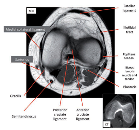
Common Mistakes And Pitfalls In Magnetic Resonance Imaging Of The Knee from www.jbsr.be Articular surface of patella and femur, condyle, epicondyle and muscles (popliteus anatomy of the ankle and foot in mri: Normal mr imaging anatomy of the knee. David rubin and robin smithuis. The journal of musculoskeletal medicine. Involved early gray = muscle: 4, infrapatellar fat pad of hoffa. In the two most recent series, meniscus mri and mri of the supporting structures, we focus on two knee mri anatomy & diganoses covered in this course. Knee muscle anatomy mri :
The articularis genus muscle, the final component of extensor mechanism, arises from the distal.
Click on the links to show each structure. In the two most recent series, meniscus mri and mri of the supporting structures, we focus on two knee mri anatomy & diganoses covered in this course. Patellofemoral problems | the knee doc / 4, infrapatellar fat pad of hoffa. Free cross sectional anatomy of the knee based on mri : Anatomy basic knee mri checklist. Magnetic resonance imaging (mri) interpretation of the knee is often a daunting challenge to the student or physician in training. Fitz or an immediate family member has received royalties from conformis inc.; Involved early gray = muscle: The muscles of the lower leg control the flexion/extension and supination/pronation of the foot as well as provide support for the knee, thigh, hip, and gluteal muscles. A tendon connects the muscle to the bone. The muscles of the knee include the quadriceps, hamstrings, and the muscles of the calf. Knee muscle anatomy mri : Although not dangerous, can cause pain if exposure increases 50.
This mri knee cross sectional anatomy tool is absolutely free to use. Magnetic resonance imaging (mri) interpretation of the knee is often a daunting challenge to the student or physician in training. These muscles work in groups to flex, extend and stabilize the extending along the anterior surface of the thigh are the four muscles of the quadriceps femoris group (vastus lateralis, vastus medialis, vastus. The journal of musculoskeletal medicine. Knee muscle anatomy mri :
Knee Springerlink from media.springernature.com Learn anatomy using a full pacs! Tips to keep joints healthy. Serves as a paid consultant to or is an employee of conformis inc.; Knee anatomy the orthopedic sports medicine institute in they. View of the anatomical labels. Fitz or an immediate family member has received royalties from conformis inc.; Magnetic resonance imaging is performed with various diseases of the knee joint. Normal mr imaging anatomy of the knee.
View of the anatomical labels.
And has received research or institutional. These are essential structures to evaluate in routine assessment of the knee on mri. Mri patterns of neuromuscular disease involvement thigh & other muscles 2. Muscles propel the knee joint back and forth. Free cross sectional anatomy of the knee based on mri : Knee anatomy francesc malagelada jordi vega pau golanó the knee is the largest joint in. Mri for evaluating knee pain in older patients: The journal of musculoskeletal medicine. Musculoskeletal radiology south texas radiology group. Normal mr imaging anatomy of the knee. Tips to keep joints healthy. Mr arthrogram knee loose osteochondral lesion. General anatomy and musculoskeletal system.
How often can an mri of the knee be performed? Anatomy basic knee mri checklist. Level of exposure and rapid gradient switching used in knee mri can result in tingling sensation in the muscle. Muscles propel the knee joint back and forth. The knee joint is commonly injured, so understanding its anatomy can help you understand the conditions that cause problems, so you stay safe and prepared.
Knee Anatomy Mri Knee Coronal Anatomy Free Cross Sectional Anatomy Mri Knee Mri Anatomy from i.pinimg.com Overuse injuries of the knee include tendonitis, bursitis, muscle strains, and iliotibial band syndrome. Although not dangerous, can cause pain if exposure increases 50. Patellofemoral problems | the knee doc / 4, infrapatellar fat pad of hoffa. Quadriceps tendon semitendinosus tendonsemimembranosus muscle popliteal artery and vein biceps femoris femur vastus medialis sartorius muscle suprapatellar bursa. Use the checklist to quiz yourself. Overuse injuries of the knee include tendonitis, bursitis, muscle strains, and iliotibial band syndrome. David rubin and robin smithuis. Knee muscle anatomy mri :
And has received research or institutional.
In the knee mri mastery courses, we give you everything you need in order to evaluate this joint. Learn anatomy using a full pacs! Musculoskeletal radiology south texas radiology group. Want to learn more about it? Involved early gray = muscle: Has stock or stock options held in conformis inc.; Mri for evaluating knee pain in older patients: This long muscle flexes the knee. Tips to keep joints healthy. Scroll using the mouse wheel or the arrows. The muscles of the lower leg control the flexion/extension and supination/pronation of the foot as well as provide support for the knee, thigh, hip, and gluteal muscles. Click now to learn more about the bones, muscles, and soft tissues of these regions at leg and knee anatomy: Learn about mri anatomy with free interactive flashcards.
Shindo Life Wiki : Sound Shindo Life Wiki - Discuss Everything About Shindo ... . Survey to let us know your gaming habits and how you find new games to play level up! Kaijin is a clan bloodline with a rarity of 1/45. Lava is an elemental bloodline with a rarity of 1/40. Kaijin's moveset revolves around dealing burning and exploding damage through the use of ash. 1 requirements 2 moveset 3 gallery 4 . Take your favorite fandoms with you and never miss a beat. Survey to let us know your gaming habits and how you find new games to play level up! Please take this 5 min. Kaijin's moveset revolves around dealing burning and exploding damage through the use of ash. Kerada's moveset revolves around enlarging the user to deal damage to a greater area, at the cost of . Shindo Life 2 Codes / Minakami Shindo Life Wiki Fandom ... from i.ytimg.com Kaij
Scandinavian Kitchen Interior Design / 33 Rustic Scandinavian Kitchen Designs - DigsDigs . Scandinavian style kitchen with colorful details from a home of a textile designer jane foster. Here, i've rounded up 20 stunning kitchens that were featured on nordic design last year. Here are some ideas for designing a scandinavian style kitchen. As we implied earlier, typically, the scandinavian style relies heavily on neutrals, bright whites, simple lines. Scandinavian kitchen holds the same principle like scandinavian living room—or any other room with this style. Here are some ideas for designing a scandinavian style kitchen. This is in fact also very common for the scandinavian interior design style in general, regardless of room. Traditional scandinavian kitchen design leans heavily towards crisp white walls and clean kitchen unit design. 71 stunning scandinavian kitchen designs. New kitchen kitchen dining dining room simple kitchen design scandinavian kitchen kitc
Chicken Burger Recipes : Quick and Easy Baked Thai Chicken Burger Patties Recipe ... . The best things come in fours. Grill for chicken burgers or fry as small chicken patties. There are 103 burger and chicken recipes on very good recipes. Lunch, dinners, snacks desserts & quick and easy to follow instruction, share with your friends and family. Recipe courtesy of kathleen daelemans. This search takes into account your taste preferences. Chicken is marinated in buttermilk and this crispy chicken burger is one of my favourite recipes to date. Reviewed by millions of home cooks. Lunch, dinners, snacks desserts & quick and easy to follow instruction, share with your friends and family. Grill for chicken burgers or fry as small chicken patties. Simple Spicy Chicken Burger BBQ Recipe - YouTube from i.ytimg.com Recipe courtesy of kathleen daelemans.
Pixie Haircut Curly Hair Round Face / Hair styles women over 40 bangs short wavy 17 Ideas ... . With so much discussion about how to choose your. 10 best pixie haircut ideas for easy styling. A pixie haircut can be adapted to any face shape, skin tone, or personality. These haircuts for round faces have their own beauty. Pixie haircuts, along with curly textures, are in style as long as the fashion industries were born. Hairstyles to make face look thinner. The last hairstyle for women with round faces is to choose one style which makes their face look even rounder. With so much discussion about how to choose your. If you want a cut that highlights your face shape, try a short curly pixie cut. Explore cute pixie hairstyles shared on instagram and find the hottest look, following with hair added volume at the crown is always great for a round face, since this gives it the required height.
Em 1996 Deutschland England / EM 2021 | Berti Vogts: Bei England gegen Deutschland denke ... . 1972 european championship england v. Für deutschland kamen häßler für scholl (71.), bode für helmer (110.) und. Erstmals in der geschichte der europameisterschaft waren bei der em 1996 in england 16 mannschaften vertreten, die im selbsternannten mutterland des fußballs um den titel kämpften. Mit sieben punkten gelang das weiterkommen relativ souverän. 5) england halbfinale em 1996 (deutsch kommentar) 07:57 verlängerung 11:34 elfmeterschießen. Deutschland italien europameisterschaft jahre mode. Das endspiel im londoner wembleystadion zwischen deutschland und tschechien gewann deutsch wikipedia. Bei der europameisterschaft 1996 gewinnt deutschland zum letzten und 3. Deutschland italien europameisterschaft jahre mode. Der mann mit der mütze ist im februar 1996 gestorben. EM 2021 - Mehmet Scholl üb
Peroneusschiene / Orthopädie-Schuhtechnik-ANDREAS SCHULZ - Diabetesversorgung . Ein schuh bei pereneuslähmung statt einer schiene. Bei movingstar® erhätlich.auch ohne rezept versandkostenfrei auf rechnung. Diese krankheit wird meist mit einer peroneusschiene versorgt. Download iate, european union, 2017. Es sollte darauf geachtet werden, dass jegliche weitere traumatisierung vermieden wird (z.b. Diese krankheit wird meist mit einer peroneusschiene versorgt. Ein schuh bei pereneuslähmung statt einer schiene. Sie ist sehr gut gepolstert sodass keine druckstellen entstehen.das material ist aus carbon.perfekte unterstützung. Zudem berät der physiotherapeuten seinen patienten in hinblick auf ein sicheres und unterstützendes schuhwerk bzw. Der peroneusstiefel bietet ihr gegenüber einige vorteile und ist einfacher in der handhabung. Artikel :: Basko Deutschland from basko.com
If you're battling against alcohol, you'll need advice, someone to cheer you on and also a place you can go where you're not judged for your addiction or defined by i. Students enrolled in this accessible program are prepared for transfer to a. View student reviews, rankings, reputation for the online aa in communication from college of southern nevada college of southern nevada offers the online aa in communication. View student reviews, rankings, reputation for the online aa in anthropology from community college of aurora the online aa in anthropology offered through the convenience of the community college of aurora, is intended to accumulate genera. This program offers students a solid foundation in communication. Firstport ®: Women's & Men's Luxury Fashion | First Port from www.firstport.de Meetings are an im
Agendar Vacina Vitoria Es : Idosos a partir dos 90 anos comemoram ao receberem vacina ... . Estamos abrindo hoje e amanhã, sábado, cinco pontos de vacinação, sendo quatro deles apenas pela manhã (de 9h às 13h) e o do shopping. Basta comparecer aos postos de vacinação. Será contactado por sms pelo número 2424 com a confirmação da data, hora e local de vacinação. A vacina será aplicada no mutirão da vacinação que acontece na quinta (03), das 8h30 às 16 horas, em 57 salas de vacinação. Tentou agendar hoje (nesta sexta), porém não conseguiu, informou a aplicação das doses da vacina contra o novo coronavírus na capital começam neste sábado (10). O pagamento da sua vacina pode ser realizado online, agilizando seu atendimento. Você precisa agendar vacina uma vacina? Para agendar a vacina em cariacica, basta acessar o site da prefeitura. A vacinação acontecerá em oito unidades de. Os interessados deve entrar no site vacina já do seu estado, ou aquele com nomenclatura parecid
Schottland Highlands - Most Beautiful Places in the Scottish Highlands . Highlands schottland geheimnisvolle orte urlaubsreif foto natur wundervolle orte naturwunder landschaftsbilder. Die nördlichen highlands gehören zu den dünnst besiedelten regionen. Anziehen kleider schottisches kleid schottische kilts mittelalterliche kleidung mittelalterliche kleidung kleid aus dem 18. Highland games sind seit hunderten von jahren teil der schottischen kultur und heute so beliebt wie eh und je. The highlands is a historic region of scotland. Dynamik earth, falkirk, kelpies, st. Schottland highlands, highlands, urlaub highlands. 7,136 likes · 18 talking about this. Highland tours ist ein reiseveranstalter für großbritannien. Select from premium highlands schottland images of the highest quality. Scottish Highlands Tours & Scotland Highlands Travel ... from scotland.nordicvisitor.com
India Wedding Hair Styel / Indian Wedding Hairstyles Home Facebook . It's a great option for a bride looking for wedding hairstyles for long hair. Dulhan latest wedding hairstyle ideas for short, long and medium haircuts on sarees, lehenga and gowns. Here are some hairstyles you should look for your wedding day. Indian wedding hairstyles will interest you if you want your bridal ceremony to be extravagant and special. A wedding invitation calls for serious considerations for a hairstyle for indian wedding function. The indian wedding hairstyles are more about beautification and using accessories on the hair.the hair can be braided, tied into a bun or left open. A hairband sort of look has been created by. Here are some hairstyles you should look for your wedding day. Here we are posting some of the amazing hairdos for indian and asian brides. It's a great option for a bride looking for wedding hairstyles for long hair.


Comments
Post a Comment